
Childhood Jaundice: from Jaundice in babies to jaundice in older children
Jaundice in children can present very differently depending on the age of the child. The approach to a newborn with jaundice is different to an older child or adolescent. One of the first and most important things is to differentiate unconjugated from conjugated hyperbilirubinemia. In neonatal life, neonatal cholestasis is particularly important as early diagnosis of conditions such as biliary atresia can significantly influence outcomes.
Neonatal cholestasis
Neonatal cholestasis is the reduced bile formation or flow resulting in the retention of biliary substances within the liver normally excreted into bile. It is characterized by jaundice with conjugated bilirubin ≥1 mg/dl, detected in a newborn or infant younger than 6 months old.
The term “neonatal hepatitis” is sometimes used in this setting, but is not a diagnosis or any distinguished entity. In earlier days, anything which was not biliary atresia was sometimes described as neonatal hepatitis. It is essentially a histological term referring to evidence of giant cell hepatitis on liver biopsy for whom no other etiology was confirmed.
The etiological spectrum of neonatal cholestasis has changed over time. Earlier descriptions included a large proportion of cases labelled as “idiopathic neonatal hepatitis”, whereas more recent evaluation has identified structural, genetic, metabolic, infectious and other causes. Common etiologies include biliary atresia, paucity of intrahepatic bile ducts, progressive familial intrahepatic cholestasis (PFIC) and metabolic liver disease. The Max CLBS 2026 data include biliary atresia/choledochal cyst, PFIC, metabolic liver disease, infections and idiopathic cases among the causes of neonatal cholestasis as mentioned in the research study published by Dr Vipul Gautam at the end of this blog.
Biliary atresia
Biliary atresia is a progressive fibro-inflammatory obliteration of the extra- and intrahepatic bile ducts that disrupts the flow of bile from the liver to the duodenum. Biliary atresia occurs worldwide and is equally distributed between males and females. The incidence mentioned in the presentation is 1:5000 in East Asia and 1:15,000 in the USA.
There are two broad types of biliary atresia: fetal/embryonic type, accounting for 10–20%, and post-natal type, accounting for 80–90%. The fetal/embryonic type is associated with BASM and presents in the immediate perinatal period.
Biliary atresia splenic malformation syndrome may be associated with splenic malformation, most commonly polysplenia. Other associated anomalies include situs inversus (37%), preduodenal portal vein (40%), absent inferior vena cava (70%), intestinal malrotation (60%), cardiac anomalies such as ventricular septal defect, atrial septal defect and hypoplastic left heart (45%), and pancreatic anomalies (11%).
Early diagnosis of biliary atresia
Early diagnosis is the key in biliary atresia. Screening can include urine and stool colour charts. A cross-sectional screening study of 1,24,385 infants born at 14 Texas hospitals between January 2015 and June 2018 evaluated a two-stage screening method. Infants were tested within the first 60 hours of life using direct bilirubin >97.5th percentile, followed by repeat testing at 2 weeks of life.
Stool colour is particularly important. In the diagnostic steps for biliary atresia, stool colour has a sensitivity of 96% and specificity of 74%. GGT has a sensitivity of 82% and specificity of 83%, while abdominal ultrasound has a sensitivity of 71% and specificity of 82%. Liver biopsy has a sensitivity of 95% and specificity of 100%, and cholangiogram has a sensitivity of 100% and specificity of 100%.
GGT can also help in the evaluation of neonatal cholestasis. Low GGT cholestasis includes the PFIC spectrum and BASD. High GGT cholestasis includes biliary atresia, Alagille syndrome, PFIC type 3, neonatal sclerosing cholangitis and TPN-associated cholestasis/multifactorial cholestasis.
Role of ultrasound in biliary atresia
Ultrasound is an important part of the evaluation. In biliary atresia, findings may include a non-visualized CBD, a small gallbladder <15 mm or a non-visualized gallbladder, the triangular cord sign with an echogenic shadow >3.5 mm, increased subcapsular flow and a prominent hepatic artery. The ultrasound should also comment on BASM.
In choledochal cyst, the presentation mentions a dilated CBD >1.5 mm in newborns and >3 mm in infants.
The role of HIDA scanning needs to be considered carefully. Acholic stools are equally informative as non-excretory HIDA. HIDA can delay the diagnosis and is only indicated when stool colour is fluctuating, to exclude biliary atresia.
Liver biopsy has >90% accuracy for biliary atresia. The important histological findings include bile ductular proliferation, portal expansion, portal and periportal fibrosis and bile plugs.
Cholangiography is another important diagnostic step. The presentation describes percutaneous cholecystocholangiography as a tool to conclusively exclude biliary atresia, with the advantage of being gold standard like intraoperative cholangiogram and potentially save the baby from negative laparotomy.
ERCP and MRCP are technically difficult in the early infantile period and have low yield. They are not included in the routine evaluation of neonatal cholestasis.
A typical clinical picture of biliary atresia can be that of a happy growing child with progressively increasing jaundice and white stools, high GGT levels and hepatosplenomegaly.
Management of biliary atresia
Kasai portoenterostomy remains an important part of the management of biliary atresia. The presentation describes Type 1, Type 2 and Type 3 biliary atresia, with Type 3 accounting for approximately 90%.
The age at Kasai has an important relationship with native liver survival. The data presented show native liver survival at 15 years of 40% when Kasai was performed at 31–45 days, 33% at 46–60 days, 28% at 61–75 days, 16% at 76–90 days and 13% when performed after 90 days.
The data from ILBS also highlight the importance of early diagnosis. In the presented cohort, 11.4% underwent diagnosis before 60 days, 50.6% between 60–90 days, 25.3% between 90–120 days and 12.7% after 120 days of life.
Progressive familial intrahepatic cholestasis
Progressive familial intrahepatic cholestasis consists of inherited diseases of hepatocellular origin resulting in disrupted bile formation. Retention of bile acids is an important part of the pathogenesis of cholestasis in PFIC. It is associated with increased risk of hepatocellular carcinoma and progression to cirrhosis and end-stage liver disease. Severe pruritus is common.
Alagille syndrome
Another cause of neonatal and childhood cholestasis is Alagille syndrome, characterized by paucity of intralobular bile ducts. The blog refers to a retrospective study of 117 children with Alagille syndrome and describes the presence of bile duct paucity and at least 3 of 5 classical criteria.
Alagille syndrome should be suspected in the context of chronic cholestatic jaundice and bile duct paucity associated with typical facial features, heart sounds (peripheral pulmonary artery stenosis), vertebral abnormalities (butterfly vertebrae), posterior embryotoxon, or renal dysfunction. Diagnosis can be confirmed by liver biopsy showing paucity of intrahepatic bile ducts and genetic analysis for pathogenic JAG1 or NOTCH2 variants. Assessment should include liver function, fat-soluble vitamins, nutritional and growth status, and cardiac and renal function.
Management is primarily supportive and aimed at controlling cholestasis, pruritus and nutritional deficiencies. Caloric intake should include high calorie feeds (including medium chain triglycerides) and supplementation for vitamins A, D, E and K. Medications for cholestatic pruritus can include ursodeoxycholic acid, rifampicin, cholestyramine or naltrexone; IBAT inhibitors such as maralixibat may be indicated. Biliary diversion may be required for resistant cases, whereas liver transplantation should be considered for progressive liver dysfunction, severe cholestasis/pruritus that is unresponsive to therapy or consequences of end-stage liver disease.
The figure attached shows an Alagille syndrome child who underwent liver transplantation for end stage liver disease. Panel 1 shows Jaundice before transplant and Panel 2 shows how child became normal after transplant.
Figure-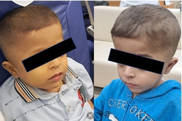
Metabolic liver disease
Metabolic liver disease includes inborn errors of metabolism which involve the liver either as hepatomegaly and/or hepatic dysfunction in the clinical presentation.
There are several important clinical clues. Galactosemia can present with early cholestasis, vomiting, hypoglycemia, E. coli sepsis and cataracts. Tyrosinemia Type I can present with acute liver failure, coagulopathy, renal Fanconi syndrome and elevated AFP. Alpha-1 antitrypsin deficiency is the most common inherited metabolic liver disease causing neonatal cholestasis, with variable severity. Niemann–Pick Disease Type C can present with cholestasis and splenomegaly, followed later by neurological manifestations. Mitochondrial hepatopathies can present with cholestasis or liver failure, lactic acidosis and multisystem involvement. Citrin deficiency (NICCD) can present with neonatal cholestasis, fatty liver, hyperammonemia and aminoacidemia.
A practical approach to neonatal cholestasis
The initial approach to neonatal cholestasis includes giving injection Vitamin-K, performing LFTs and checking the colour of three consecutive stools. Acholic or fluctuating stool colour should prompt fasting ultrasound and early referral for possible biliary atresia.
The clinical context is also important. Prematurity, TPN usage, a sick child, synthetic failure, PFIC, Niemann-Pick C, metabolic liver disease, galactosemia, tyrosinemia, infections such as sepsis and UTI, and choledochal cyst are among the possibilities that need to be considered. In specific scenarios, CMV, HSV, ferritin, AFP and urine succinylacetone may be required.
The blog also provides a practical list of common treatable causes of neonatal cholestasis and the relevant investigations. For biliary atresia, GGT levels, ultrasound abdomen and liver biopsy are listed. For choledochal cyst, ultrasound abdomen is the investigation. Galactosemia can be evaluated with urine routine, urine for reducing substances and serum GALT assay. Tyrosinemia can be evaluated with serum AFP and urine succinylacetone. Sepsis or multifactorial cholestasis requires a sepsis screen including urine routine. Endocrine causes can be evaluated with FT4/TSH levels, fasting blood sugar and serum cortisol, while BASD can be evaluated with bile acids and GGT.
Jaundice in older children
The approach to jaundice in older children begins by differentiating conjugated from unconjugated hyperbilirubinemia.
Unconjugated hyperbilirubinemia is related to increased bilirubin production or impaired conjugation. Urine is normally coloured, without bilirubinuria. Stool is generally normal and pruritus is absent. Liver enzymes are usually normal. Common causes include hemolytic anemia such as G6PD deficiency, hereditary spherocytosis and autoimmune hemolysis, Gilbert syndrome, Crigler–Najjar syndrome and ineffective erythropoiesis.
Conjugated hyperbilirubinemia is related to hepatocellular dysfunction or impaired bile flow. Urine is dark because bilirubinuria is present, stool may be pale or clay-coloured in cholestasis, and pruritus is common in cholestatic disorders. Liver enzymes are often abnormal, with elevated ALT/AST or ALP/GGT.
The common causes listed include acute viral hepatitis such as HAV, HEV and HBV, autoimmune hepatitis, Wilson disease, drug-induced liver injury, gallstones or choledocholithiasis, primary sclerosing cholangitis and choledochal cyst. Initial evaluation includes CBC, reticulocyte count, peripheral smear, LDH, haptoglobin and Coombs test for unconjugated hyperbilirubinemia, while evaluation of conjugated hyperbilirubinemia includes LFTs, PT/INR, GGT, abdominal ultrasound, viral markers, autoimmune profile and Wilson work-up.
Dark urine in a jaundiced child should be considered conjugated hyperbilirubinemia until proven otherwise.
Acute liver Failure and acute hepatitis
Acute liver injury or acute hepatitis in children can have a wide range of causes. Viral infections include HAV, HBV, HEV, EBV, CMV, HSV and adenovirus. Enteric fever, atypical infections and dengue fever can also be associated.
Drug- and toxin-induced causes include paracetamol, anti-tubercular drugs, antiepileptics, herbal medications and mushroom poisoning. Autoimmune hepatitis is another important cause. Ischemic and vascular causes include dehydration, sepsis or shock and Budd–Chiari syndrome. Other causes include hemophagocytic lymphohistiocytosis (HLH), malignant infiltration and indeterminate disease where the etiology remains unknown.
Pediatric acute liver failure is an important clinical entity that requires early recognition. Causes include infections such as hepatitis A, E and B, EBV, CMV, Parvo, dengue, VZV and HSV. Metabolic causes include Wilson disease and infantile liver failure due to tyrosinemia, galactosemia, urea cycle disorders and mitochondrial disease. Autoimmune liver disease and drug- or toxin-induced liver injury are also important causes.
Drugs and toxins listed in the presentation include paracetamol, ATT drugs, valproate and other antiepileptics, amoxycillin-clavulanic acid, erythromycin/azithromycin, sulfa drugs, quinolones and herbal medicines.
Chronic liver disease
Chronic liver disease refers to disease of the liver which lasts over a period of six months. It is a progressive disorder characterized by ongoing hepatic injury leading to fibrosis and architectural distortion, with or without cirrhosis, and is not solely defined by duration of illness.
It is important to understand the difference between chronic liver disease, cirrhosis, chronic hepatitis and acute on chronic liver failure. Chronic liver disease is progressive deterioration of liver functions over six months. Cirrhosis is varying degrees of fibrosis replacing the healthy parenchyma, secondary to long-standing damage and inflammation. Chronic hepatitis is ongoing inflammation within the liver that is capable of progression to cirrhosis and liver failure. Acute on chronic liver failure occurs when an acute insult occurs in a diseased liver, previously known or not, and causes liver decompensation, organ failures and poor outcome.
Chronic liver disease can be in an early asymptomatic stage, referred to as compensated chronic liver disease, or a later symptomatic stage, referred to as decompensated chronic liver disease. Decompensation is characterized by the appearance of ascites, hepatic encephalopathy and gastrointestinal bleeding.
Children with chronic liver disease are not always symptomatic. When symptoms are present, they may be related to the liver parenchyma or to portal hypertension.
Causes of chronic liver disease according to age
In children between 1–5 years, important causes include biliary atresia after Kasai surgery, autoimmune hepatitis, PFIC with late presentation, Alagille syndrome with late diagnosis, metabolic liver disease and choledochal cyst.
In children older than 5 years and adolescents, important causes include Wilson disease, autoimmune hepatitis, primary sclerosing cholangitis with or without inflammatory bowel disease, chronic HBV/HCV infection, choledochal cyst and PFIC-like type 3 disease.
Wilson disease
Wilson disease usually presents after 3 years of age and can present with hepatitis, cirrhosis or acute liver failure. Neurological symptoms may occur in older children, and Coombs-negative hemolytic anemia can be an important clinical clue.
Management includes copper chelation with D-penicillamine or trientine, zinc therapy and liver transplantation for liver failure or decompensated cirrhosis.
Autoimmune hepatitis
Autoimmune hepatitis predominantly affects girls and adolescents. It may present with recurrent jaundice, with or without itching, and can be asymptomatic or present as acute or chronic disease. Other autoimmune diseases may also be present.
Diagnosis includes elevated AST/ALT, raised serum IgG and ANA, ASMA or Anti-LKM positivity. Liver biopsy confirms the diagnosis. Management includes corticosteroids and azathioprine/MMF for maintenance, with long-term follow-up.
Drug-induced liver injury
Drug-induced liver injury should always be considered when there is a recent history of medication or herbal product use. The presentation can be variable, with either hepatocellular or cholestatic injury. Common drugs include ATT, antiepileptics and antibiotics.
Diagnosis requires a careful drug history and exclusion of other causes. Improvement after withdrawal supports the diagnosis. Management includes stopping the offending drug, supportive care and monitoring liver function. If liver failure develops, referral is required.
Primary sclerosing cholangitis
Primary sclerosing cholangitis can present with persistent cholestatic jaundice, fatigue, fever and pruritus. It may be associated with inflammatory bowel disease and hepatosplenomegaly.
Diagnosis includes cholestatic liver enzymes with increased GGT, MRCP showing multifocal biliary strictures and liver biopsy. Management includes symptomatic treatment and drugs as modifiers, endoscopic management of dominant strictures and endoscopy to rule out ulcerative colitis. Liver transplantation may be required for advanced disease.
Choledochal cyst and choledocholithiasis
Choledochal cyst or choledocholithiasis can be incidental or present with intermittent jaundice, right upper quadrant pain and fever if cholangitis is present.
Ultrasound abdomen is the first-line investigation, while MRCP is used for anatomy and obstruction. Management includes antibiotics for cholangitis, definitive surgical excision for choledochal cyst and ERCP or surgery for choledocholithiasis.
Jaundice in children: the overall approach
Jaundice in children should not be approached as a diagnosis; it is just a symptom of some disease. The age of the child, duration of jaundice, clinical history, examination and pattern of liver injury all help narrow the diagnosis.
In neonatal cholestasis, the priority is to differentiate conjugated hyperbilirubinemia and identify biliary atresia early. Stool colour, GGT, ultrasound, liver biopsy and cholangiography all have roles in the diagnostic pathway. Other important causes include PFIC, Alagille syndrome, metabolic liver disease, infections, endocrine disorders, choledochal cyst and multifactorial cholestasis.
In older children, the first step is to distinguish unconjugated from conjugated hyperbilirubinemia. If conjugated hyperbilirubinemia is present, the next step is to determine whether the liver disease is acute or chronic and whether the pattern is predominantly hepatocellular or cholestatic.
The red flags that should prompt urgent attention include acute liver failure, coagulopathy, encephalopathy and portal hypertension. These children should be referred promptly to a pediatric hepatology or transplant centre.
Interestingly, the CLBS data presented in the blog show that biliary atresia remains an important diagnosis even beyond infancy. In the presented cohort of liver transplantation, biliary atresia accounted for 142 cases (40%) at a median age of 24 months, and 71 children (50% of the biliary atresia group) had undergone Kasai portoenterostomy.
The overall message is straightforward: differentiate unconjugated from conjugated hyperbilirubinemia at the outset. Neonatal cholestasis is a medical emergency, and early diagnosis of biliary atresia is critical for improving outcomes. In older children, first determine whether the liver disease is acute or chronic and then identify the pattern of liver injury, whether hepatocellular or cholestatic.
Age, duration, history, examination and targeted investigations remain the cornerstones of diagnosis. Recognizing red flags such as acute liver failure, coagulopathy, encephalopathy and portal hypertension is essential, with prompt referral to a pediatric hepatology or transplant centre when required. Pediatric liver transplantation has excellent short- and long-term outcomes in children.
References as cited in the blog:
- De Bruyne et al. Eur J Pediatr. 2011.
- NASPGHAN, ESPGHAN. 2004.
- NASPGHAN, ESPGHAN. 2017.
- Balistreri et al. Clin Liver Dis. 2006.
- J.L. Santos et al. Brazilian Journal of Medical and Biological Research. 2010.
- Lee WS et al. Singapore Med J. 2010.
- Consensus Report on Neonatal Cholestasis. Indian Pediatrics. 2000.
- Balistreri WF. Neonatal Cholestasis. Semin Liver Dis. 1987.
- Hartley et al. Lancet. 2009.
- Yachha et al. IJG. 2020.
- Harpavat et al. JAMA. 2020.
- Clin Gastroenterol Hepatol. 2011;9:357–62e2.
- Ashritha A, Vipul Gautam, Bikrant Bihari Lal, Amar Mukund, Priti Vijay, Rajeev Khanna, Vikrant Sood, Seema Alam. Percutaneous Cholecystocholangiography—A Tool to Conclusively Exclude Biliary Atresia. Indian Journal of Pediatrics . November 2022;89(11):1144–1147.
- Serinet et al. Pediatrics. 2009;123:1280.
- Jagadisan et al. Indian J Pediatr. 2017;84:591–596.
- Kumar et al. ILBS. JCEH. 2019.
- Suchy. 5 th edition.
- JPGN. 2011.
- Alam et al. Indian J Pediatr. 2016.
- Bhatia V et al. Indian Pediatr. 2014;51.
- Squires RH Jr, Shneider BL, Bucuvalas J, et al. Acute liver failure in children: the first 348 patients in the Pediatric Acute Liver Failure Study Group. J Pediatr . 2006;148(5):652–658.
- Alam et al. Liver International. 2017.
- APASL – Asia Pacific Association for the Study of Liver Disease.
- EASL – European Association for the Study of the Liver.
Original Research Studies conducted by Dr. Vipul Gautam in Childhood Jaundice and Cholestatic Liver Disease
- Vipul Gautam has contributed to the understanding of genetic causes of cholestasis in children, including rare forms of PFIC. In a 2024 case report, Dr. Vipul Gautam and colleagues reported the youngest child with KIF12-related cholestatic disease requiring liver transplantation. The 6-month-old boy had presented with neonatal jaundice, progressive abdominal distension, pruritus and hepatic decompensation. The case highlights an important diagnostic point: KIF12-related disease can present with high GGT cholestasis, making it difficult to distinguish from biliary atresia and Alagille syndrome. Whole-exome sequencing identified a homozygous KIF12 mutation, and the child subsequently underwent successful living donor liver transplantation. The report emphasizes the importance of considering genetic testing early in infants with unexplained cholestasis and timely referral when medical treatment is unsuccessful.
Citation: Gautam V, Panda K, Kumar V, Agarwal S, Gupta S. Youngest Living Donor Liver Transplant for End-Stage Liver Disease in a 6-Month-Old With a Novel Aggressive Mutation in KIF12 Gene. Pediatric Transplantation. 2024;28:e14804. doi:10.1111/petr.14804.
- Vipul Gautam, along with Rajeev Khanna, also authored a detailed review on progressive familial intrahepatic cholestasis (PFIC), an important cause of persistent jaundice and cholestasis in children. The review describes PFIC as a heterogeneous group of autosomal recessive disorders of biliary transporters, with manifestations ranging from neonatal cholestasis and recurrent jaundice to severe pruritus, growth failure, portal hypertension and end-stage liver disease requiring transplantation. The article discusses the different genetic forms of PFIC, their clinical presentation, diagnosis, pathophysiology and management, highlighting the growing role of genetic classification in guiding treatment and prognosis.
Citation: Khanna R, Gautam V. Progressive familial intrahepatic cholestasis. Pediatric Gastroenterology, Liver and Nutrition Journal (PGLJ). April–June 2020;2(2).
- In another major study, Vipul Gautam and colleagues evaluated the long-term outcomes of children undergoing living donor liver transplantation for end-stage liver disease, many of whom had presented with childhood cholestatic disorders. In this single-centre study, 480 pediatric liver transplants were performed, with 448 children surviving beyond one year. Biliary atresia was the most common indication, accounting for 40% of cases, followed by PFIC at 10%, metabolic liver disease at 10%, acute liver failure at 8%, Wilson disease at 7.5% and autoimmune hepatitis at 7%. Among the 358 children included in the long-term analysis, the median follow-up was 62 months, and survival probabilities were 95%, 93%, 91% and 72.4% at 3, 5, 10 and 15 years, respectively. The study provides long-term outcome data from a large Indian pediatric living-donor liver transplant program and demonstrates the importance of timely management of children progressing from cholestatic liver disease to end-stage liver disease.
Citation: Gautam V, Nekarakanti PK, Kumar V, Das DJ, Bashir S, Singh SA, Gupta S. Long-term Prognosis in Pediatric Living Donor Liver Transplant Recipients Who Have Survived the First Year of Surgery. Journal of Clinical and Experimental Hepatology. 2025;15(6):102631. doi:10.1016/j.jceh.2025.102631.
For more information on childhood jaundice and parents’ counselling, Dr Vipul Gautam is routinely available at Vasant kunj Clinic and Max Hospital, Saket, New Delhi.
About the Author
Dr. Vipul Gautam, MD, DM
Consultant Pediatric Hepatologist & Pediatric Liver Transplant Physician
Centre for Liver and Biliary Sciences, Max Super Speciality Hospital, Saket, New Delhi
Private Clinic: Vasant Kunj, New Delhi
Clinic timings: Monday–Saturday, 7:00 AM–9:00 AM and 7:00 PM–9:00 PM
Sunday: By appointment
Areas of clinical interest: Pediatric gastroenterology, hepatology, neonatal cholestasis, biliary atresia and pediatric liver transplantation.
Last updated: 02-October-2026